Vital 3 Joint Solution Clinical Trials
Full Text of Harvard Study. Effects of Oral Administration of Type II Collagen on Rheumatoid Arthritis
Study performed by David E. Trentham*, Roselynn A. Dynesius-Trentham, E. John Orav, Daniel Combitchi, Carlos Lorenzo, Kathryn Lea Sewell, David A. Hafler, Howard L. Weiner
12 Week Randomized, Double-Blinded, Placebo Controlled, Parallel Group Study (AI-200-002). Published in the prestigious journal Science September 24, 1993; 261:1727-1730
Copyright ©1993 by the American Association for the Advancement of Science
Abstract
Rheumatoid arthritis is an inflammatory synovial disease thought to involve T cells reacting to an antigen within the joint. Type II collagen is the major protein in articular cartilage and is a potential autoantigen in this disease. Oral tolerization to autoantigens suppresses animal models of T cell-mediated autoimmune disease, including two models of rheumatoid arthritis. In this randomized, double-blind trial involving 60 patients with severe, active rheumatoid arthritis, a decrease in the number of swollen joints and tender joints occurred in subjects fed chicken type II collagen for 3 months but not in those that received a placebo. Four patients in the collagen group had complete remission of the disease. No side effects were evident. These data demonstrate clinical efficacy of an oral tolerization approach for rheumatoid arthritis.
Article
Rheumatoid arthritis (RA) is a common chronic illness in which the synovial membrane of multiple joints becomes inflamed, causing damage to cartilage and bone. Although the pathogenetic mechanisms underlying the disease are unknown, rheumatoid arthritis is associated with human lymphocyte antigen (HSA)-DR4 and considered to be an autoimmune disorder in which activated T cells participate (1). Type II collagen is a candidate autoantigen for this disease because it is the most abundant structural protein of cartilage, and immunization of animals with the native protein creates arthritis morphologically resembling rheumatoid arthritis (2,3). Patients with the disease have immune responses to native type II collagen (4), but whether collagen reactivity participates in the primary pathogenesis of rheumatoid arthritis or reflects tissue degradation is unknown.
Current treatments are inadequate in that they only partially control established rheumatoid arthritis. They also have side effects that limit use early in the disease process and interfere with prolonged administration (5). An ideal therapy would decrease inflammation in the joint by a disease-specific mechanism and would lack toxicity. Oral tolerization, a method of inducing antigen-specific tolerance, suppresses animal models of the autoimmune diseases multiple sclerosis, uveitis, and diabetes (6-11). In a double-blind pilot trial involving 30 patients with multiple sclerosis, oral administration of bovine myelin antigens decreased the number of T cells that reacted with myelin basic protein (MBP), with no measurable toxicity (12). Although favorable trends occurred in the myelin group, clinical efficacy could not be determined because of the small sample size.
Oral administration of native type II collagen ameliorates two animal models of rheumatoid arthritis induced by type II collagen (13) or complete Freund's adjuvant (14). These experimental findings provided the rationale for a pilot, open-label dose-escalation and safety study in 10 patients with recalcitrant rheumatoid arthritis. Subjects were taken off their immuno- suppressive and disease-modifying drugs consisting of methotrexate, 6-mercaptopurine, azathioprine, or auranofin and few 0.1 mg of solubilized type II collagen daily for 1 month and then switched to 0.5 mg for the next 2 months (15). This dose was extrapolated from experiments in the rat adjuvant arthritis model where feeding 3 to 30 p.g of collagen attenuated disease (14) and the rat experimental autoimmune encephalomyelitis (EAE) model where feeding 500 to 1000 p.g of MBP was suppressive (6,10). Six of the 10 patients experienced a substantial clinical response, defined by a >50% [morning stiffness, 15-m walk time, grip strength, Westergren erythrocyte sedimentation rate (ESR), or physician or patient global assessments] and lasting for at least 2 months after the treatment period (16). A complete response, that is, disease remission (17) with discontinuation of nonsteroidal anti-inflammatory drug (NSAID), occurred in one patient previously on methotrexate and continued for 26 months. There were no adverse effects. Based on the results of this phase I study, a placebo-controlled, phase II trial was undertaken to determine whether clinical efficacy could be demonstrated.
For this phase II trial, 60 patients with severe, active rheumatoid arthritis and who met eligibility criteria (18) gave informed consent (19) and were entered into the study. They were withdrawn from immuno-suppressive drugs if they had been taking them (20) and randomized (21) to either a treatment identical to that used in the phase I trial (15) or an indistinguishable placebo (22) to be taken orally for a consecutive 90-day period. Both patients and investigators, except those responsible for medication (23), were masked as to treatment. Assessments were performed by the same investigator (D.E.T.) At the initiation of treatment and a 1,2, and 3, months, generally at the same time of day (24).
At the conclusion of the study, 59 of the 60 patients were considered evaluable (25); 28 had received collagen and 31 placebo. On entry, demographic, clinical, and laboratory parameters were similar in both groups (Table 1) (26). Relative to entry, there was significant (P >0.05) improvement in the number of swollen joints, the number of tender or painful joints, joint-swelling and tenderness indices, and 15-m walk time at months 1, 2, and 3 in the colagen group as compared with placebo patients, except for the number of tender or painful joints at month 2 (P = 0.06) (Table 2). Among the collagen patients (14%), as compared with none in the placebo group, had complete resolution of disease (27). Table 3 indicates the patients' status by other outcome measures (16, 21, 28). Stability or improvement while patients were off immunosuppressives occurred in the collagen group, whereas patients in the placebo group tended to deteriorate. In alternative analyses that reduce the influence of the four placebo patients who withdrew from the trial (25), a similar significant (P >0.05) improvement from collagen was seen (29). A placebo effect resembling that encountered in other RA trials (30) was also observed. Four patients (13%) in the placebo group exhibited substantial benefit (16) and attained functional class I ranking. This observation reaffirms the critical importance of placebo-controlled evaluations in rheumatoid arthritis. No side effects or significant changes in laboratory values, including rheumatoid factor and antibodies to type II collagen, were noted. There was no evidence of sensitization to collagen, as measured by antibodies to type II collagen. Attempts to assess T cell responses to type II collagen, including release of transforming growth factor-B (TGF-8), were unsuccessful because of the difficulty in demonstrating reactivity to type II collagen in the peripheral blood of RA patients. None of the baseline features, including the presence of collagen antibodies, HLA haplotype, or sex, were associated with responsiveness to collagen (31).
This controlled trial provides evidence that oral administration of small quantities of solubilized native heterologous type II collagen is both safe and can improve the clinical manifestations of active rheumatoid arthritis. Baseline values were determined while 64% of the collagen-treated patients were on immunosuppressive drugs (usually methotrexate or 6-merca[topurine), and further improvement occurred with collagen treatment. If longer term efficacy is established, oral collagen would be a preferable treatment because it is not toxic. Although it is possible that the disease could be exacerbated or an allergy to the oral antigen could develop, this was not observed in our study, in animals(6-11, 13, 14), in multiple sclerosis patients given oral myelin for as long as 3 years (12), or in uveitis patients treated with retinal S-antigen (32). All patients in the phase II trial and open-label trial had collagen discontinued after 3 months. Four patients in the pilot study who improved while on collagen experienced a relapse about 3 months after cessation of therapy followed by benefit with reinitiation of collagen. In animals, protective effects of oral tolerance appear to last for 2 to 3 months after termination of antigen feeding (6). Recrudescence of disease after discontinuation of oral toleragen has also occurred in multiple sclerosis (12) and uveitis (32) patients. It therefore appears that additional administration is required to maintain the clinical effects of oral tolerance.
On the basis of studies of oral tolerance in animals, two immunologic mechanisms could explain the clinical response to collagen observed in this study. Feeding type II collagen in RA cases may both anergize CD4+ type II collagen autoreactive cells and generate major histocompatibility complex (MHC) class I- or class II- restricted regulatory cells that sequester within joint tissues and release cytokines that inactivate autoaggressive cells. In animals, feeding large doses of antigen favors T cell anergy, whereas multiple small doses favors the induction of regulatory T cells (33). In the EAE model, feeding low doses of MBP activates MBP-specific regulatory cells in gut lymphoid tissue (10). These cells are predominantly CD8+ and suppress EAE by trafficking to the central nervous system and releasing anti-inflammatory cytokines, such as TGF-8 and interleukin-r, when they encounter MBP presented by MHC molecules in inflamed brain tissue. This process, termed antigen-driven bystander suppression (10), implies that an orally administered protein can down-regulate organ-specific autoimmune disease as long as it is a constituent of the target tissue and is capable of inducing regulatory I cells. It is not obligatory for the protein to have the disease-inciting epitopes. Examples of bystander suppression include inhibition of proteolipid protein (PLP) - induced EAE by orally administered MBP (34), delay of diabetes in the non-obese diabetic mouse by oral insulin (11), and abrogation of adjuvant arthritis by oral collagen (14). In all three models, autoimmunity to the toleragen does not appear to initiate disease. Accordingly, our data do not determine whither type II collagen is the primary autoantigen in rheumatoid arthritis.
Although initial clinical efficacy of oral collagen has been shown, questions concerning optimum dosing and long-term control of disease remain. Nonetheless, this study demonstrates the therapeutic efficacy of oral tolerance for a human autoimmune disease and provides the foundation for the development of oral collagen as an easily administered nontoxic treatment for rheumatoid arthritis.
Table 1. Patient Characteristics
Patient characteristics at entry. There were no differences between groups (P>0.10) detected by either Fisher's exact test or the Wilcoxon rank-sum test (age and disease duration).
| Characteristic | Collagen Treatment (n=28) | Placebo Treatment (n=31) |
|---|---|---|
| Age (years=SD) | 50.3 = 11.9 | 55.1 = 12.9 |
| Sex (% females) | 71 | 68 |
| Disease duration (years=SD) | 9.8 = 6.2 | 10.3 = 8.1 |
| Rheumatoid factor [%,(number tested)] | 74 (27) | 82 (28) |
| HLA-DR 4+ [%,(number tested)] | 45 (28) | 62 (29) |
| Collagen II antibody [%, titer greater than or equal to 2) | 32 | 13 |
| Prednisone (%, less than or equal to 10 mg/day) | 25 | 48 |
| Immunosuppressive* withdrawn (%) | 64 | 58 |
*Methotrexate, 6-mercaptopurine, azathioprine, hydroxychloroquine, sulfasalazine, auranofin, cyclosporin, cyclophosphamide, or penicillamine. Seven patients were receiving combinations of these drugs (20). The remaining patients were not on immunosuppressive drugs at the time of entry because of prior lack of response to toxicity to at least two of the drugs.
Table 2. Disease Variables
Disease variables in collagen- versus placebo-treated patients (collagen/placebo) evaluated at entry = 28/31, 1 month = 27/29, 2 months = 26/26, and 3 months = 28/31; withdrawals were treated as described (25); values shown are different from entry except for patient and physician assessments which are given as percentages. There were no significant differences between groups at entry (P > 0.05 for all variables by the Wilcoxon rank-sum test or the x2 trend test for patient and physician assessments) (16). Comparisons between groups showed significantly more improvement or less worsening in the collagen-treated patients (P < 0.05 and P < 0.01).
Differences between physician assessments in collagen and placebo patients were not significant but showed trends in favor of collagen at 1 month (P = 0.066 and 2 months (P = 0.06). Qualitatively similar results were found when a two-way analysis of variance was used to adjust for prednisone use. Significant improvement was also observed among collagen-treated patients at 1, 2, and 3 months in terms of the number of swollen joints, the swollen joint index, the number of tender joints, and the tenderness index (Student's t test; all P values are <0.01, except at 3 months, for the number of swollen joints, P = 0.02, and the swollen joint index, P = 0.03).
| Variable | Group | Mean value at entry (=SE) | Change from entry at month 1 | Change from entry at month 2 | Change from entry at month 3 |
|---|---|---|---|---|---|
| Joints Swollen (number) | Collagen | 11.8 = 0.9 | -2.7 = 0.5** | -4.1 = 1.0* | -3.1 = 1.1* |
| Joints Swollen (number) | Placebo | 12.0 = 0.8 | 2.0 = 1.4 | 0.9 = 1.6 | 1.3 = 1.4 |
| Joints tender to pressure or painful on passive motion (number) | Collagen | 15.8 = 1.3 | -4.1 = 1.1* | -6.7 = 1.5 | -5.4 = 1.8* |
| Joints tender to pressure or painful on passive motion (number) | Placebo | 15.6 = 0.8 | 1.1 = 1.4 | -1.1 = 1.7 | -0.1 = 1.6 |
| Joint-Swelling Index | Collagen | 13.3 = 1.1 | -3.4 = 0.8** | -4.8 = 1.2* | -3.1 = 1.4* |
| Joint-Swelling Index | Placebo | 13.2 = 0.9 | 2.4 = 1.8 | 0.9 = 1.6 | 4.3 = 2.1 |
| Joint-tenderness or pain index | Collagen | 17.5 = 1.3 | -5.0 = 1.2** | -7.6 = 1.7* | -5.7 = 2.0* |
| Joint tenderness or pain index | Placebo | 17.2 = 1.0 | 1.6 = 1.8 | -0.5 = 2.1 | 3.0 = 2.4 |
| 15-m walk time (s) | Collagen | 13.2 = 0.6 | 0.0 = 0.3** | 0.25 = 0.5** | 0.5 = 0.6** |
| 15-m walk time (s) | Placebo | 14.9 = 0.9 | 1.9 = 0.6 | 3.8 = 1.2 | 20.8 = 7.5 |
| Grip Strength (mmHG) | |||||
| Right | Collagen | 105 = 9 | 0.1 = 6.0 | 6.3 = 7.8 | -0.9 = 8.5 |
| Right | Placebo | 87 = 8 | -7.3 = 6.2 | -8.3 = 8.4 | -16.4 = 8.8 |
| Left | Collagen | 106 = 10 | 0.6 = 5.6 | 6.6 = 7.4* | -0.3 = 8.8 |
| Left | Placebo | 95 = 8 | -8.9 = 5.8 | -9.3 = 10.1 | -13.8 = 9.7 |
| Morning stiffness duration (min) | Collagen | 155 = 51 | 64.8 = 106 | 51.2 = 100 | 56.4 = 92 |
| Morning stiffness duration (min) | Placebo | 210 = 55 | 130 = 76 | 168 = 108 | 195 = 100 |
| Patient assessment (%) | |||||
| Absent or mild | Collagen | 21 | 41 | 23* | 36* |
| Moderate | Collagen | 54 | 33 | 46* | 25* |
| Severe or very severe | Collagen | 25 | 26 | 31* | 39* |
| Absent or mild | Placebo | 16 | 21 | 15 | 19 |
| Moderate | Placebo | 35 | 31 | 23 | 10 |
| Severe or very severe | Placebo | 48 | 48 | 62 | 71 |
| Physician Assessment | |||||
| Absent or mild | Collagen | 18 | 41 | 35 | 32 |
| Moderate | Collagen | 46 | 33 | 38 | 29 |
| Severe to very severe | Collagen | 36 | 26 | 27 | 39 |
| Absent or mild | Placebo | 6 | 21 | 27 | 19 |
| Moderate | Placebo | 42 | 31 | 12 | 13 |
| Severe to very severe | Placebo | 52 | 48 | 62 | 68 |
| ESR (mm/hour) | Collagen | 39 = 6 | 5.1 = 2.9 | 4.9 = 2.8 | 1.7 = 3.9 |
| ESR (mm/hour) | Placebo | 34 = 5 | 9.8 = 5.0 | 7.8 = 5.6 | 3.2 = 2.8 |
*P<0.5
**P<0.01
Table 3. Outcome Measures
Outcome measures in collagen- versus placebo-treated patients. Values are percentages of 28 collagen and 31 placebo patients.
| Variable | Entry Collagen | Entry Placebo | Three months Collagen | Three months Placebo |
|---|---|---|---|---|
| Worsening status* | 7* | 35 | ||
| Analgesic use† | 14† | 39 | ||
| Functional class†† | ||||
| I | 0 | 0 | 18 | 13 |
| II | 57 | 58 | 39 | 19 |
| III | 43 | 42 | 39 | 59 |
| IV | 0 | 0 | 4 | 10 |
*Represents an increase of 30% or more from the entry value for the joint-swelling index and the joint-tenderness or pain index (16). Comparison between groups showed significantly more deterioration in the placebo-treated patients (P is less than or equal to 0.01 by the Fisher's exact test.)
**Narcotic without anti-inflammatory properties, usually acetaminophen with codeine, propoxyphene, or pentazocine, prescribed at any time by the clinical investigator in an attempt to retain flaring patients in the trial. Comparison between groups showed significantly greater numbers of placebo treated patients requiring narcotics (P < 0.04 by the Fisher's exact test).
***Determined by American Rheumatism Association criteria for functional class (28) I, no limitation from arthritis; II, mildly restricted; III, Markedly restricted, and IV, incapacity causing virtual bed or wheelchair existence. Trend for improvement in the collagen group not significant (P = 0.10 by the x2 trend test.
References & Notes
- K.L. Sewell and D.E. Trentham, Lancet 341. 283 (1993).
- D.E. Trentham, A.S. Townes, A.H. Kang, J. Exp. Med. 146, 857, (1977).
- J.S. Courtenay, M.J. Dallman, A.D. Dayan, A. Martin, B. Mosedale Nature 283, 666 (1980); E.S. Cathcart et al., Lab. Invest. 54, 26 (1986).
- N.A. Andriopoulos et al., Arthritis Rheum. 19, 613 (1976); D.E. Trentham, R.A. Dynesius, R.E. Rocklin, J.R. David, N Engl. J. Med. 299, 327 (1978); A. Tarkowski, L. Klareskog, H. Carlsten, P. Herberts, W.J. Koopman, Arthritis Rheum. 32, 1087 (1989).
- P.M. Brooks, Lancet 341, 286 (1993).
- P.J. Higgins and H.L. Wiener, J. Immunol. 140, 440 (1988).
- D. Bitar and C.C. Whitacre, Cell. Immunol. 112, 364 (1988); C.C. Whitacre, I.E. Gienapp, C.G. Orosz, D. Bitar, J. Immunol. 147, 215 (1991).
- R.B. Nussenblatt et al, J. Immunol. 144, 1689 (1990).
- O. Lider, M.B. Santos, C.S.Y. Lee, P.J. Higgins, H.L. Weiner, ibid. 142, 748 (1989).
- A. Miller, O. Lider, H.L. Weiner J. Exp. Med. 174, 791 (1991); S.J. Khoury, W.W. Hancock, H.L. Weiner, ibid. 176, 1355 (1992); A. Miller, O. Lider, A. Roberts, M.B. Sporn, H.L. Weiner, Proc. Natl. Acad. Sci. U.S.A. 89, 421 (1992).
- Z.J. Zhang, L. Davidson, G. Eisenbarth, H.S. Weiner, Proc. Natl. Acad. Sci. U.S.A. 88, 10252 (1991).
- H.L. Weiner et al., Science 259, 1321 (1993).
- C. Nagler-anderson, L.A. Bober, M.E. Robinson, G.W. Siskind, G.J. Thorbecke, Proc. Natl. Acad. Sci. U.S.A. 83, 7443 (1986); h.s.g. Thompson and N.A. Staines, Clin. Exp. Immunol. 64, 581 (1986).
- Z.J. Zhang, C.S.Y. Lee, O. Lider, H.L. Weiner, J. Immunol. 145, 2489 (1990).
- Native type II collage, isolated from sternal cartilage of chicks rendered lathyritic by administration of 8-aminopropionitrile (2), was used to treat the first rive subjects in the phase I pilot study. Subsequent patients in the pilot trial and in the double-blind study received type II collagen purified from non arthritic chicken sternal cartilage by the identical technique (2) and obtained from Genzyme (Boston, MA). Preparations were analyzed for purity by standard biochemical methods (2, 35) and tested for arthritogenicity and toxicity in rats (2) with findings of batch-to-batch equivalency. Collagen was stored in a lyophilized state (2) at -20C with desiccant. The protein was solubilized in 0.1 M acetic acid for -12 hours at 4 C, sterilized by membrane filtration, and aliquoted into individual 1.0-ml doses in sterile tubes. Tubes sufficient for about 2 weeks of treatment were delivered on ice to patients and maintained under refrigeration until use. For oral administration, the 1.0-ml aliquot was added to 4 to 6 ounces (118 to 177 ml) of cold orange juice and the mixture drunk immediately. Orange juice provided an additional acid vehicle to inhibit precipitation of collagen and masked the taste of acetic acid. All dosing occurred in the morning on an empty stomach at least 20 min before breakfast or ingestion of other fluids. Smoking was not permitted during this interval.
- M.E. Weinblatt et al., N. Engl. J. Med. 312, 818 (1985); K.L. Sewell et al., Arthritis Rheum., in press.
- R.S. Pinals, A.T. Masi, R.A. Larsen, Arthritis Rheum, 24, 1308(1981).
- The following requirements determined eligibility: (i) American Rheumatism Association (ARA) criteria for classic or definite rheumatoid arthritis (16); (ii) onset of the disease at age 16 or older; (iii) age of at least 18 years; (iv) ARA functional class unresponsive to at least one immunosuppressive (Table 1); and (vi) severe active disease defined by at least three of the following: at least nine painful or tender joints, at least six swollen joints, at least 45 min of morning stiffness, or at least 28 mm/hour ESR. Exclusion criteria included a degree of structural joint damage not amenable to physical rehabilitation if inflammation subsided after treatment or a serious concurrent medical problem. Some patients (n=39) represented referrals for treatment of refractory disease by rheumatologists outside Boston; others (n-10) had received experimental therapy for rheumatoid arthritis in the past.
- The study was approved by the Beth Israel Hospital Committee on Clinical Investigations and conducted under an investigator-initiated Investigational New Drug (IND) permit from the U.S. Food and Drug Administration.
- Because of the possibility that patients would receive ineffective therapy or a placebo, study medication was begun immediately after the patient discontinued immunosuppressive drugs (Table 1); patients receiving parenteral gold were not entered because prolonged carryover effects could influence the outcome. Patients remained on their NSAID, prednisone dose (less than or equal to 10 mg/day), or both, during the 3-month treatment period. NSAID substitution, increases in NSAID or prednisone dose, or initiation of any other antirheumatic therapy with the exception of analgesic agents and intraarticular steroids represented protocol violations. If applicable, patients were requested to practice contraception.
- A biostatistician (E.J.O.) randomized each patient to either the active or placebo treatment group in blocks of six, stratified by functional class (28) severity.
- The placebo consisted of 1.0-ml doses of 0.1 M acetic acid subjected to membrane filtration.
- Three investigators (D.C., C.L., and K.L.S.) obtained the randomization and prepared medication but did not have access to clinical data. No unblinding occurred.
- Conventional instruments were used to measure RA activity (16). Assistive devices were permitted for walk times. The clinical investigator cared for the patients during the trial and was responsible for safety monitoring. Laboratory safety assessment was performed immediately before randomization and at 2, 4, 8, and 12 weeks thereafter. The assessment comprised a complete blood count, differential and platelet count, liver and renal function tests, prothrombin and partial thromboplastin times, urinalysis, and ESR. HLA typing was performed for the alleles of the A,B, C and DR/DQ loci (36). Serum immunoglobulin M (IgM) rheumatoid factor titers were determined by nephelometry and IgG antibody titers to native type II collagen (expressed as -log2) by enzyme-linked immunosorbent assay (37) immediately before and at the end of collagen or placebo administration.
- Before unbinding, decisions were made concerning the analysis of five subjects (8%) that failed to complete the study. One was noncompliant and withdrew for personal reasons on day 40 after only a baseline examination. This patient was excluded from analysis and had been randomized to collagen. Four discontinued their study medication before the end of the 3-month treatment because of worsening arthritis. They were assigned the worst score in the sample for the remainder of the study and included in the analysis. All four had been randomized to placebo. One protocol violation occurred with a patient who increased the daily dose of prednisone from 5 mg to 10 mg just before month 2. Because the patient continued to do poorly and the 10-mg dose was consistent with eligibility requirements, the patient was included in the analyses; the patient had been randomized to collagen. No steroid injections or other problems with compliance occurred.
- Comparisons between collagen- and placebo-treated patients were performed with the Wilcoxon rank-sum test for continuous measures (such as the number of swollen joints), the Fisher's exact test for dichotomous measures (such as narcotic usage), and the x2 trend test for functional class and patient and physician assessments. All measured end points such as the number of swollen joints were compared with entry values before testing; qualitative measures, such as patient and physician assessments and functional class, are presented and analyzed without adjustment for baseline responses. The Student's paired t test was used to assess whether changes in the collagen group represented significant improvements over baseline values. Reported P values were two-sided.
- Complete resolution is a more rigorous extension of RA remission criteria (17), preformulated because of the magnitude of improvement in some patients in the initial trial, and is defined by the following conditions: no swollen or tender joints, no morning stiffness or afternoon fatigue, absent arthritis on physician and patient appraisals, functional class I status, and normal ESR (<28 mm/hour) while off prednisone.
- O. Steinbrocker, C.H. Traeger, R.C. Batterman, J. Am. Med. Assoc. 140, 659 (1949).
- Rather than assigning the placebo patients who withdrew from the trial the worst observed value (25), they were given the value from their last visit. Because one of the four dropped out before the 1-month follow-up, that patient was removed from all analyses, reducing the sample size to 28 collagen and 30 placebo patients. By this analysis, the number of tender joints, joint-tenderness index, walk time, patient assessment of severe or very severe disease, and analgesic use was significantly (P is less than or equal to 0.05) improved in the collagen group compared with the placebo group.
- H.J. Williams et al., Arthritis Rhuem. 31, 702 (1988).
- Analysis of variance showed no significant interaction between treatment effectiveness (as measured by changes in the number of swollen joints, tender joints, or walk time) and any characteristic in Table 1.
- R. Nussenblatt, personal communication.
- A. Friedman and H.L. Weiner, J. Immunol. 150, 4A (1993): H.L. Weiner et al., Ann. Rev. Immunol., in press.
- A. Al-Sabbagh, A. Miller, R.A. Sorbel, H.L. Weiner, Neurology 42 (suppl.3), 346 (1992).
- S.M. Helfgott, R.A. Dynesius-Trentham, E. Brahn, D.E. Trenthma, J. Exp. Med. 162, 1531 (1985).
- G.M. Kammer and D.E. Trentham, Arthritis Rheum. 27, 489 (1984).
- S.M. Helfgott et al., Lancet 337, 387 (1991).
- Supported by NIH grants MO1 RRO1032 and AG00294 and by a grant from Autoimmune Inc. We thank E. Milford and C.B. Carpenter for HLA-typing. In accordance with disclosure guidelines of the Harvard Medical School, D.A.H. and H.L.W. have a financial interest in Autoimmune Inc.
- Trentham DE, Dynesius-Trentham RA, Orav EJ, et al. Effects of Oral Administration of Type II Collagen on Rheumatoid Arthritis. Science 1993; 261:1727-1730.
Vital 3 Joint Solution® Products
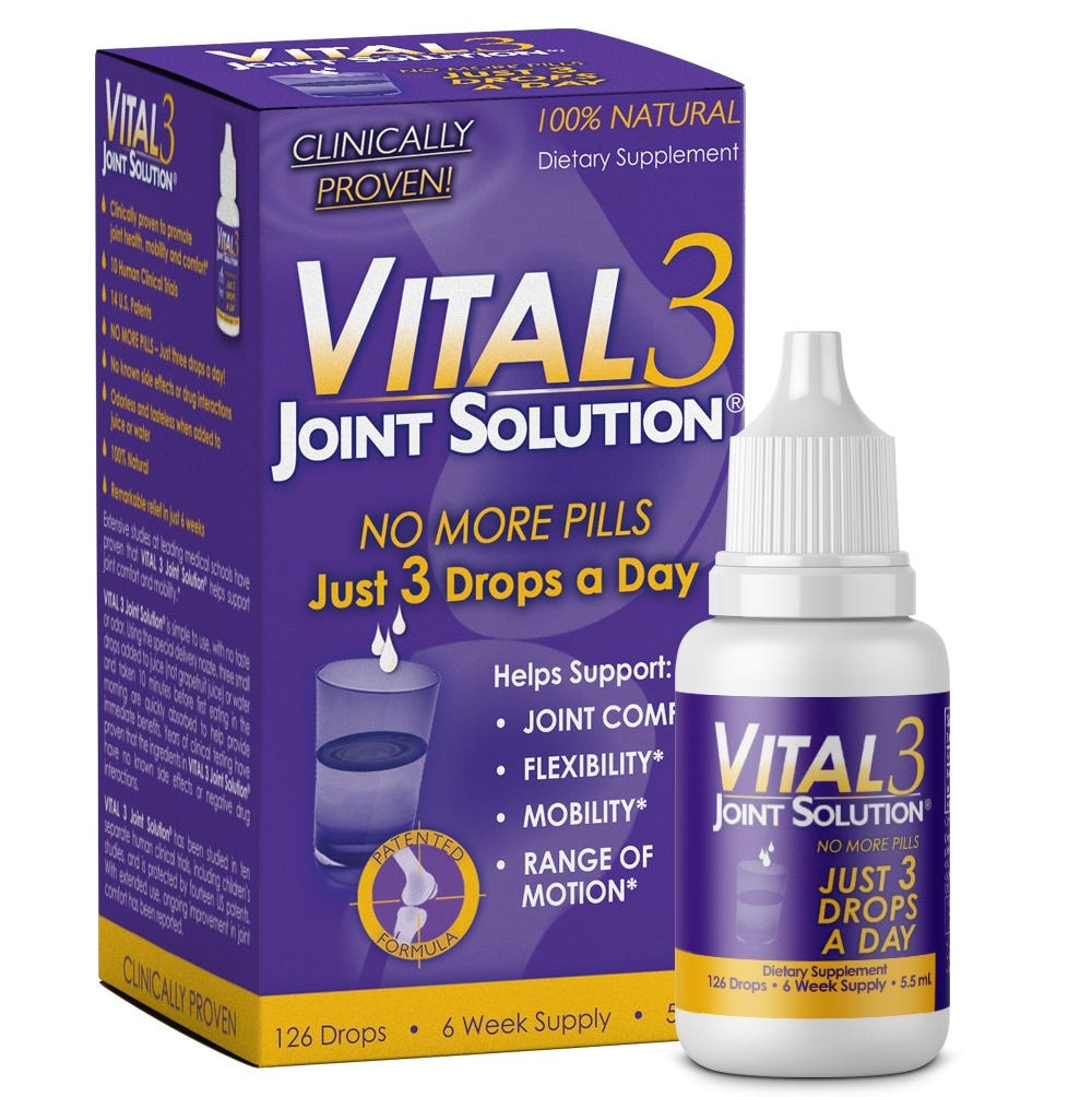
Vital 3 Joint Solution® - 5.5 mL
Your Price: $38.99
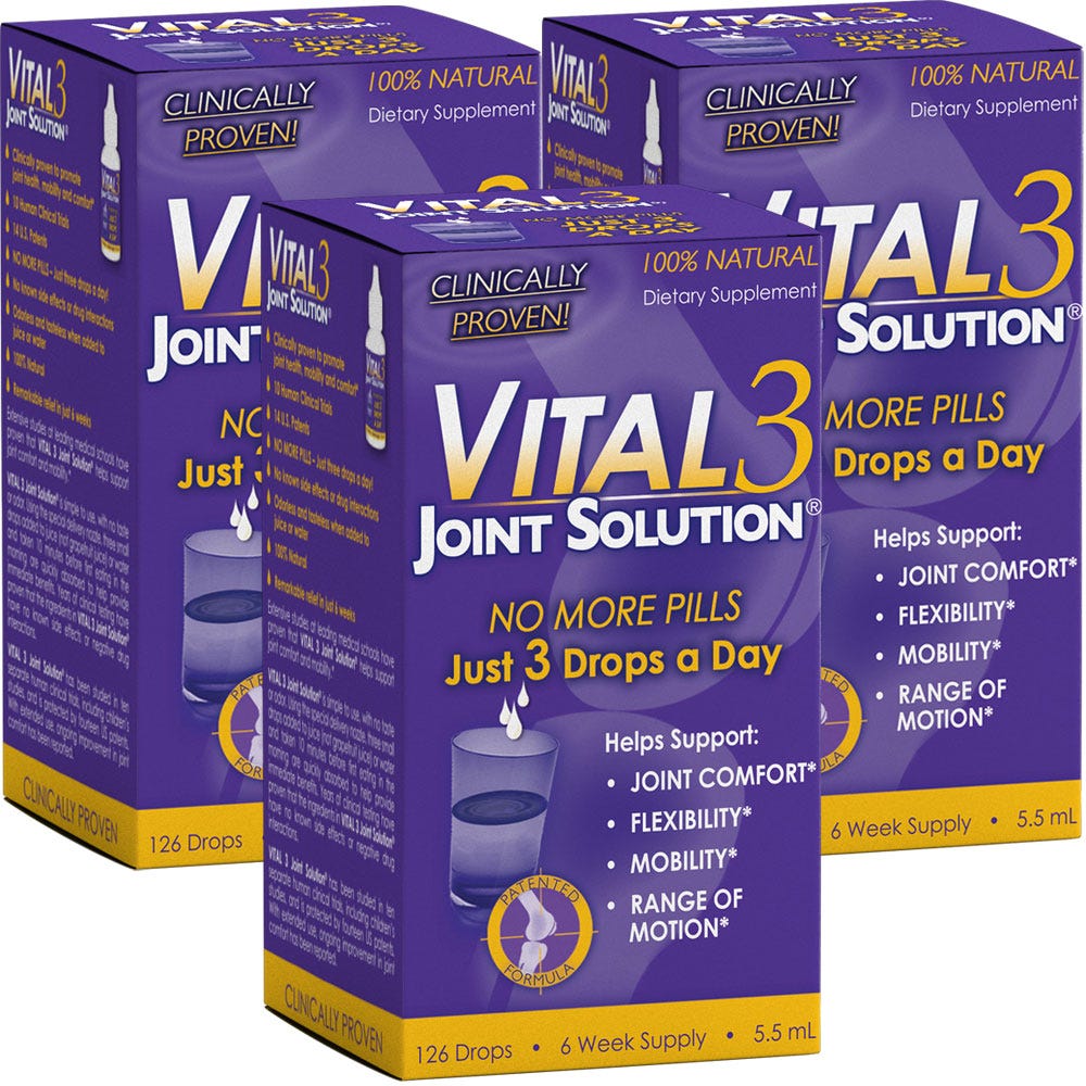
Vital 3 Joint Solution® - 16.5 mL (3 Bottles)